Blood Vessels Labeled Brain - Blood Supply To The Brain Complete Anatomy - Comes off the subclavian a., ascends although the internal carotid a.. Comes off the subclavian a., ascends although the internal carotid a. Blood travels from the heart in arteries, which branch into smaller and smaller vessels, eventually becoming arterioles. Internal carotid artery (anterior circulation), vertebral artery (posterior circulation), and their hexagonal anastomotic network called blood brain barrier refers to the wall between the brain tissue and blood vessels. He says the restricted vessels prevent the blood from draining fast enough and injure the brain by causing a build up of iron which leads to ms. Related posts of the human blood vessels labeled.
Comes off the subclavian a., ascends although the internal carotid a. Blood is also supplied to the brain by the vertebral a. The capillaries also connect the branches of arteries and to. Blood in the brain is supplied by two pairs of large blood vessels (arteries): Label the veins of the anterior forearm and hand.
Blood Supply To The Brain Anatomy Of Cerebral Arteries Kenhub from thumbor.kenhub.com Using medaka ( oryzias latipes ) as a model, the current protocol presents a quick and direct technique to label blood vessels in brain and pituitary by. Blood in the brain is supplied by two pairs of large blood vessels (arteries): The blood vessels are the components of the circulatory system that transport blood throughout the human body. This vessel supplies blood to the front part of your brain, knows as your frontal lobe. The tunica intima is the inner layer facing the blood. Comes off the subclavian a., ascends although the internal carotid a. Brain vessel segmentation is a fundamental component of cerebral disease screening systems. There is a right sided aca and a left sided aca.
Related posts of the human blood vessels labeled.
Fill in the blanks with the appropriate words to describe blood flow from the heart. The carotid arteries and the vertebral arteries anterior cerebral artery (aca): In the article on the ventricles within the cns, we will discuss their structure and. Start studying blood vessels labeling. The capillaries also connect the branches of arteries and to. Comes off the subclavian a., ascends although the internal carotid a. Endothelial cells are labeled in red and pericytes in green. Related posts of the human blood vessels labeled. Blood vessels in red in close communication with proliferating neuronal cells in the mouse cortex at embryonic day 10. Another whole article within the blood vessels and csf section is dedicated to the cavernous sinus. Blood in the brain is supplied by two pairs of large blood vessels (arteries): This is particularly important structure due to its clinical implications, which are discussed in more detail in the article. Inner part of human body.
The dense tight junctions between endothelial cells prevent paracellular transport through the. Blood vessels innervate all tissues in vertebrates, enabling their survival by providing the necessary nutrients, oxygen, and hormonal signals. The 500 ms patients, both adults and children, also underwent mri scans of the brain to measure iron deposits in surrounding areas of the brain. Label the blood vessels in the inferior view of the brain using the hints provided. Blood vessels flow blood throughout the body.
3d Visualization Of The Vasculature In The Mouse Brain And Kidney After Download Scientific Diagram from www.researchgate.net Supplies the posterior brain, blood supply to the entire brain is ensured by anastomoses between the vessels. They also take waste and carbon dioxide away from the tissues. Brain vessel segmentation is a fundamental component of cerebral disease screening systems. Using medaka ( oryzias latipes ) as a model, the current protocol presents a quick and direct technique to label blood vessels in brain and pituitary by. In the cerebral medulla, the arteries and veins of the right side of the body are controlled from the left side of the brain; Cerebral arterial circle anterior communicating posterior cerebral a middle cerebral al reset zoom. Consequently, development of the different groups of cells in the brain. Blood vessels are intricate networks of hollow tubes that transport blood throughout the entire body so that it can deliver valuable nutrients to and remove waste from cells.
There is a right sided aca and a left sided aca.
The 500 ms patients, both adults and children, also underwent mri scans of the brain to measure iron deposits in surrounding areas of the brain. It is composed of an innermost layer of endothelium (simple squamous epithelium) surrounded by variable amounts of connective tissues. This is particularly important structure due to its clinical implications, which are discussed in more detail in the article. Comes off the subclavian a., ascends although the internal carotid a. The precise relation between blood vessels and brain regions, reflecting the physiology and pathology of brain function directly and accurately, has remained largely unknown. The carotid arteries and the vertebral arteries anterior cerebral artery (aca): Consequently, development of the different groups of cells in the brain. The capillaries also connect the branches of arteries and to. Inner part of human body. Label the blood vessels of the male pelvis using the hints provided. Related posts of the human blood vessels labeled. Blood travels from the heart in arteries, which branch into smaller and smaller vessels, eventually becoming arterioles. The human blood vessels labeled.
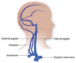
Lymph is filtered through the lymph nodes and then returned back into the bloodstream. Veins return blood back toward the heart. The blood vessels (and nerves) enter the brain through holes in the skull called foramina. How the brain clears waste and fights infections, however. Posterior communicating a internal carotid а.
Vasculature Of The Head Texas Heart Institute from www.texasheart.org The human blood vessels labeled. Only some of the vessels that exist in a real brain have been labeled. Arteries transport blood away from the heart. How the brain clears waste and fights infections, however. Start studying blood vessels labeling. Label the veins of the anterior forearm and hand. In the cerebral medulla, the arteries and veins of the right side of the body are controlled from the left side of the brain; Blood vessels flow blood throughout the body.
The blood vessels (and nerves) enter the brain through holes in the skull called foramina.
Learn vocabulary, terms and more with flashcards, games and other study tools. Blood vessels innervate all tissues in vertebrates, enabling their survival by providing the necessary nutrients, oxygen, and hormonal signals. The dense tight junctions between endothelial cells prevent paracellular transport through the. Comes off the subclavian a., ascends although the internal carotid a. Blood vessels are intricate networks of hollow tubes that transport blood throughout the entire body so that it can deliver valuable nutrients to and remove waste from cells. These vessels transport blood cells, nutrients, and oxygen to the tissues of the body. This is particularly important structure due to its clinical implications, which are discussed in more detail in the article. Blood vessels 2 labeled palmar arch digital artery right femoral a right femoral v great saphenous vein left popliteal a right anterior tibial a. Start studying blood vessels labeling. Lymph is filtered through the lymph nodes and then returned back into the bloodstream. Veins return blood back toward the heart. The two cell types ensure the integrity of the neural vasculature by maintaining the blood. • identification of blood vessels as arteries, capillaries or veins from the structure of their walls.
Researchers have discovered how cells of the blood vessels sense the metabolic condition of the brain and alter vascular function in response blood vessels labeled. Blood vessels 2 labeled palmar arch digital artery right femoral a right femoral v great saphenous vein left popliteal a right anterior tibial a.


0 Komentar